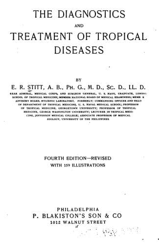
The diagnostics and treatment of tropical diseasesStitt, E. R. (Edward Rhodes)
Science
The diagnostics and treatment of tropical diseases
Stitt, E. R. (Edward Rhodes)
Tropical medicine
Natives of India very rarely develop liver abscess but it has been
noted that when they begin to follow the customs of Europeans, as
to eating and drinking, such lesions become more common in them.
As to the proportion of cases of amoebic dysentery which give
rise to liver abscess only the statistics of those who have
differentiated between bacillary and amoebic dysentery are of any
value. Such statistics would indicate that about 20% of the cases
of amoebic dysentery are complicated by liver abscess.
Liver abscess may be present without demonstrable lesions in the
large intestines, such lesions having healed or the intestinal
involvement having been so slight as not to have caused other than
microscopic changes.
It is a well-known fact that liver abscess may set in years after a
patient has left the tropics and years after the occurrence of any
dysenteric manifestations.
PATHOLOGY
There seems little doubt but that the amoebae in the thrombosed
terminals of the portal vein are carried by way of the upward current
into the liver where they lodge in the liver capillaries, Councilman
and Lafleur having found amoebae in such emboli.
Another view is that the amoebae may wander across the abdominal
cavity and enter the liver in this way. This seems as improbable as
that view which considers a possible entrance by way of the bile
duct. Bile is toxic to amoebae and it would be difficult to explain
their presence in the small intestines.
In 639 cases Roux found the abscess in the right lobe in 70% of the
cases.
Other statistics give about 75% for the right lobe, 10% for the
left lobe, 4% for the lobus Spigelii and in about 10% of cases
abscesses are found in both right and left lobes.
In 562 cases Zancarol found a single abscess in 60% of the cases.
In 288 cases Waring found a single abscess in 61.5%, double abscesses
in 11.5% and multiple abscesses in 27%. The favorite site of liver
abscess is the superior and posterior part of the right lobe and near
its surface.
The abscesses vary enormously in size, some being no larger
than a walnut while others may contain a quart or more of pus,
exceptionally as much as a gallon. The pus is typically of a
chocolate color and contains degenerated liver cells, granular
débris and often haematoidin and Charcot-Leyden crystals. There is
an absence of polymorphonuclears. It may however be creamy in color.
In Strong’s cases about 50% of the abscesses showed bacteria upon
culturing, the organisms noted being staphylococci, streptococci,
_B. coli_ and _B. pyocyaneus_.
The walls of liver abscesses are rather shaggy and the amoebae are
found deeply located.
It is probable that the necrotic process, set up by the amoebae,
begins in the interlobular capillaries although it may at times
begin within the lobule.
Public-domain text, read in full here on John Shaqi.
Reviews
Reviews
No reviews yet
Be the first to share your thoughts on this work.
Elsewhere in the archive
Join the Discussion
Join the discussion
Sign in to leave a comment or review.
Sign InorCreate an account