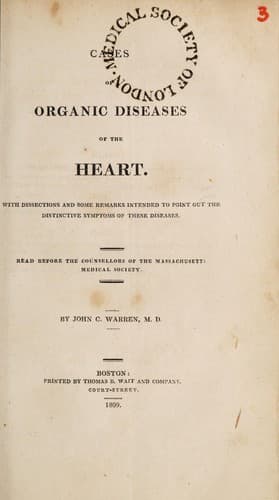
Cases of Organic Diseases of the Heart — John Shaqi
Cases of Organic Diseases of the HeartWarren, John Collins
Science
Cases of Organic Diseases of the Heart
Warren, John Collins
Heart -- Diseases
The cartilages of the ribs were ossified. The left cavity of the
pleura contained about twelve ounces of water; the right, about three
ounces. The lungs, externally, were dark coloured, especially the
posterior lobes; internally, they were very firm, and, in some places,
as dense as the substance of the liver. A frothy mucus was effused
from them in great quantities. They were coloured by very dark blood,
especially in the middle portion of the left superior lobe. One or two
calcareous concretions were observed in them. The pericardium was a
little firmer than usual, and contained about five ounces of water.
The heart was enlarged, and covered with tough fat. In the right
auricle, and ventricle, was some coagulated blood. The tricuspid
valves had lost their smoothness and transparency; the semilunar
valves of the pulmonary artery were cartilaginous at their bases. The
left auricle and ventricle, particularly the first, contained
coagulum. The mitral valves were roughened by many bony spots.
Considerable ossification had taken place in the semilunar valves of
the aorta, so that one of them had quite lost its form; and the aorta
was ossified for the space of a square inch, at a small distance from
the valves. The coronary arteries were also ossified.
ABDOMEN.
The coat of the liver was somewhat wrinkled, as if shrunk. Its
substance was hard, and discharged, when cut, great quantities of
blood. The veins of the omentum, mesentery, and intestines, were full
of blood. The abdomen contained a considerable quantity of water.
HEAD.
Water was found between the dura and pia mater, and between the pia
mater and arachnoides. The vertical portion of the pia mater bore
marks of former inflammation. The convolutions of the brain were very
distinct; their external surface was pale. The veins were empty[11].
No bloody points were observed in the medullary portion of the brain,
when cut. The ventricles contained between one and two ounces of
water; the communication between them was very large. The plexus
choroides was pale.
[Footnote 11: In this case, and in case first, the vena cava
ascendens had been divided, before the brain was examined.]
CASE IX.
Public-domain text, read in full here on John Shaqi.
Reviews
Reviews
No reviews yet
Be the first to share your thoughts on this work.
Join the Discussion
Join the discussion
Sign in to leave a comment or review.
Sign InorCreate an account