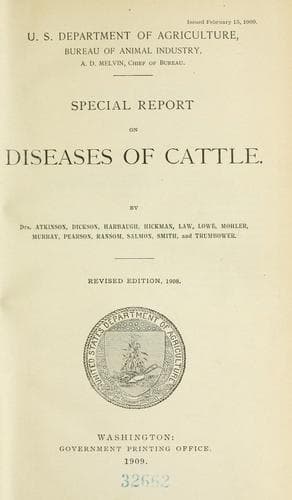
Special report on diseases of cattleUnited States. Bureau of Animal Industry
Science
Special report on diseases of cattle
United States. Bureau of Animal Industry
Cattle -- Diseases
When bleeding is performed without proper care or with an unclean lancet,
inflammation of the vein may result, or it may be caused by the animal
rubbing the wound against some object. When inflammation follows the
operation, the coats of the vein become so much enlarged that the vessel
may be felt hard and knotted beneath the skin, and pressure produces pain.
A thin, watery discharge, tinged with blood, issues from the wound. The
blood becomes coagulated in the vessel. In inflammation of the jugular the
coagulation extends from the wound upward to the first large branch.
Abscesses may form along the course of the vein. The inflammation is
followed by obliteration of that part in which coagulation exists. This is
of small import, as cattle have an accessory jugular vein which gradually
enlarges and accommodates itself to the increased quantity of blood it must
carry.
_Treatment._--The treatment for inflammation of the vein is to clip the
hair from along the course of the affected vessel and apply a blister, the
cerate of cantharides. Abscesses should be opened as soon as they form,
because there is a possibility of the pus getting into the circulation.
In the operation of bleeding the instruments should be clean and free from
rust. If the skin is not sufficiently opened, or when closing the wound the
skin is drawn out too much, blood may accumulate in the tissue, and if it
does it should be removed by pressing absorbent cotton or a sponge on the
part. Care should also be used in opening the vein, so that the instrument
may not pass entirely through both sides of the vein and open the artery
beneath it.
* * * * *
DISEASES OF THE HEART, BLOOD VESSELS, AND LYMPHATICS.
DESCRIPTION OF PLATE.
PLATE VII:
Diagram illustrating the circulation of the blood. The arrows indicate the
direction in which the blood flows. The valves of the heart, situated
between the right auricle and ventricle, and left auricle and ventricle,
and between the ventricles and large arteries, are represented by curved
lines. These valves are intended to prevent the flow of blood in a
direction contrary to that indicated by the arrows.
[Illustration: PLATE VII. DIAGRAM OF THE CIRCULATION OF THE BLOOD.]
* * * * *
NONCONTAGIOUS DISEASES OF THE ORGANS
OF RESPIRATION.
By WILLIAM HERBERT LOWE, D. V. S.
DIAGNOSIS.
Public-domain text, read in full here on John Shaqi.
Reviews
Reviews
No reviews yet
Be the first to share your thoughts on this work.
Elsewhere in the archive
Join the Discussion
Join the discussion
Sign in to leave a comment or review.
Sign InorCreate an account