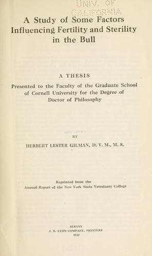
A study of some factors influencing fertility and sterility in the bullGilman, Herbert Lester
Science
A study of some factors influencing fertility and sterility in the bull
Gilman, Herbert Lester
Cattle; Infertility in animals; Livestock; Thesis (Ph. D.)
SPERMATOZOA: The history of the discovery of spermatozoa is very
interesting, and for that reason a brief outline will be given. The
“semen threads” were first observed in the year 1667, by Ham, a student
of Leeuwenhoek at Leyden. The discovery was announced, confirmed by
findings in the dog and rabbit, and discussed by the latter author under
the title: Observationes Anthonii L. de natis e semine genitali
animaculis (Upon the formation of young from procreative material). The
sperms were taken to be animals on account of their motility, and their
significance remained questionable if not unknown. Spallanzani, quoted
by Marshall (29), was the first to show that the filtered fluid was
impotent, and that spermatozoa in the semen were essential to
fertilization. Kolliker, in 1841, discovered that the sperms arise from
the cells of the testis, and Barry in 1843, observed the conjugation of
sperm and ovum in the rabbit. This led to a clear understanding of the
function of these important germinal elements.
The spermatozoa are the male procreative cells, and are characterized by
the possession of a head containing the chromosomes necessary for
fertilization, and a tail capable of propelling the organism on its way
to meet the ovum. The length of the entire sperm, including the head, is
seventy-five to eighty microns. The head is nine and five-tenths microns
long, and five and five-tenths microns wide. It may be divided into two
principal parts, the head and tail. The head, for the larger part, is
made up of the nucleus, and may be differentiated by staining reactions
into a darker staining posterior part, an anterior lighter part, and
often a still lighter area between the two. On the anterior part is a
sharpened edge, the acrosome, which serves to perforate the ovum. The
whole is surrounded by a very definite limiting membrane which often
becomes obscured under abnormal conditions. The tail may be divided into
three parts: connecting piece, principal part, and terminal filament.
The connecting piece, the essential motile apparatus, is the thickest
and strongest part, and joins the tail proper to the head. It consists
of the central axial filament, a spiral filament around this, and an
outer mitochondrial covering. Anteriorly it is limited by the anterior
portion of the posterior chromosome, and posteriorly by the annular
chromosome. The anterior chromosome is directly connected with the head,
there appearing a light break here at the neck where separation
frequently occurs. This neck serves as a joint for the motion. The axial
filament, therefore, does not reach the head, but extends back from the
anterior part of the posterior chromosome. The principal part consists
merely of the axial filament, and a thin outer covering, while the end
piece is quite thin and is made up solely of the uncovered axial
filament. The finer structures are seen only when special staining
reactions are used, and then only when the sperms are obtained directly
Public-domain text, read in full here on John Shaqi.
Reviews
Reviews
No reviews yet
Be the first to share your thoughts on this work.
Join the Discussion
Join the discussion
Sign in to leave a comment or review.
Sign InorCreate an account