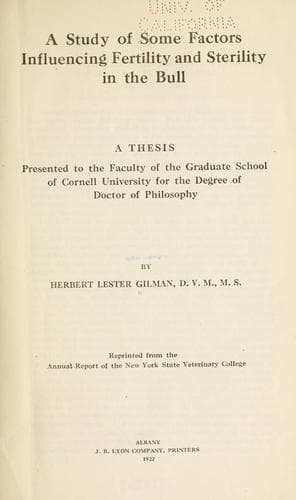
A study of some factors influencing fertility and sterility in the bullGilman, Herbert Lester
Science
A study of some factors influencing fertility and sterility in the bull
Gilman, Herbert Lester
Cattle; Infertility in animals; Livestock; Thesis (Ph. D.)
EPIDIDYMIS: This organ not infrequently presents gross abnormalities,
and very often is pathological on microscopic examination. Acute,
inflammation, with induration or abscess formation, is very common in
the tail, but less so in the head and body. Possibly this is caused by
the fact that the tail is the most pendant portion of the organ. In
these cases, the tail is enlarged, soft, and quite hot and painful on
physical examination. Enlargement, due to a connective tissue
induration, occurs occasionally in all three parts, and the inflammation
may produce adhesions to the adjacent serous membranes. Inflammation of
both the parietal and visceral layers of the tunica vaginalis is very
common. In those cases, the membrane usually is quite hyperaemic, and on
the surface it presents many small reddened tufts of newly-formed
connective tissue. In adult bulls it is exceptional not to find at least
slight evidence of some previous inflammation. In all of the numerous
bulls examined, both apparently normal and sterile, I have found but one
in which some evidence of inflammation (either present or past) could
not be found. Along with the fibrous tufts, are numerous fine strands of
connective tissues passing from the covering of the tail of the
epididymis to the adjacent portion of the parietal layer of the tunica.
The strands often extend even to the upper part of the head.
Microscopically, inflammation of the part is shown by hyperaemia, loss
of cilia of the lining cells, and exudation. In the more severe forms,
the lining cells which furnish considerable secretion for the
nourishment and stimulation of the sperms, become degenerated, and are
exfoliated into the lumen, as in Fig. 21. This condition is very common
in sterile bulls, and those of lowered fertility. In the chronic types,
the interstitial connective tissue is increased in amount, leading to
degeneration and atrophy of part or all of the tubules, as in the case
of Bull 2. Infiltration with leucocytes, and necrosis, are the
predominating lesions in the pyogenic types of inflammation.
DUCTUS DEFERENS: This tube seems to be peculiarly free from severe
inflammatory processes, and when these appear they are limited to the
mucosa. The cells of the lining membrane not infrequently show a mild
type of degeneration and exfoliation, or in the more chronic forms, the
entire membrane degenerates and disappears. In man, the duct
occasionally becomes occluded, but so far I have failed to find this
condition in the bull. Undoubtedly, when the occlusion does occur, it is
near the origin of the duct at the tail of the epididymis.
Public-domain text, read in full here on John Shaqi.
Reviews
Reviews
No reviews yet
Be the first to share your thoughts on this work.
Join the Discussion
Join the discussion
Sign in to leave a comment or review.
Sign InorCreate an account