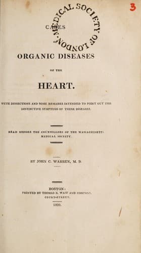
Cases of Organic Diseases of the Heart — John Shaqi
Cases of Organic Diseases of the HeartWarren, John Collins
Science
Cases of Organic Diseases of the Heart
Warren, John Collins
Heart -- Diseases
The lungs were contracted into a smaller compass than usual, and were
very firm to the touch. Their colour anteriorly was whitish, with
small distinct purple spots; posteriorly, of a deep red, with similar
spots. The right lobe adhered closely to the pericardium; it also
adhered to the pleura costalis, by a great number of strong cords,
which seemed to be elongations of the original adhesions. Some of them
were nearly as hard as ligament, and many an inch in length.
Internally the lungs presented a very compact structure. Their cells
were crowded with mucus, and their vessels filled with black blood,
partly fluid, and partly coagulated. Some portions were firmer and
more condensed than others, but no tubercles were discovered.
The pericardium, viewed externally, appeared very large, and occupied
almost the whole space behind the opening formed by removing the
sternum and cartilages of the ribs. It was situated principally on the
left side, and contained about double the usual quantity of water; but
was principally filled by the enlarged heart, to which it adhered
anteriorly about two inches, near its base. Its parietes were, in
every part, very much thickened and hardened.
The heart presented nearly its usual colour and form, excepting on its
anterior surface, which was somewhat discoloured by coagulated lymph.
It was enlarged in bulk to, at least, one half more than the healthy
size. The auricles and ventricles contained coagulated blood. The
tricuspid valves were in a sound state. The left auricle was double
the usual size. The left ventricle was enlarged, about three times
thicker and much firmer than usual. The mitral valves were very much
thickened, and near the insertion of their columnæ, which were sound,
cartilaginous, so that they were quite rigid, and the opening made by
them, from the auricle to the ventricle, was scarcely large enough to
admit the passage of a finger. The semilunar valves of the aorta were
ossified at their bases and apices, and the portion intermediate,
between the base and apex, partly ossified, and partly cartilaginous,
so as to render the valves very rigid. The aorta was at least one half
larger than usual, especially at its arch. The arteria innominata, the
carotid, and subclavian arteries, were uncommonly large and thick. The
coronary arteries were considerably ossified.
ABDOMEN.
The omentum was destitute of fat. The stomach distended with flatus on
the pyloric side; its cardiac extremity, lying under the liver, was
pressed down and contracted. The liver was shrunk; its tunic
corrugated, as if it had been distended, and bearing marks of
inflammation; its substance harder than usual; its vessels, when
divided, pouring out liquid black blood. The gall bladder was filled
with bile. The kidneys were thicker, and more irregular in form, than
is common. The abdominal cavity contained some water.
HEAD.
Public-domain text, read in full here on John Shaqi.
Reviews
Reviews
No reviews yet
Be the first to share your thoughts on this work.
Join the Discussion
Join the discussion
Sign in to leave a comment or review.
Sign InorCreate an account