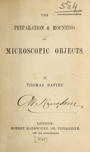
The preparation & mounting of microscopic objectsDavies, Thomas
Science
The preparation & mounting of microscopic objects
Davies, Thomas
Microscopy -- Technique
NERVE-TISSUE.--This is seldom mounted; as Dr. Carpenter observes, “no
method of preserving the nerve-tissue has been devised which makes it
worth while to mount preparations for the sake of displaying its minute
characters,” but we will mention a few particulars to be observed in
its treatment. The nerve should be taken from the animal as soon as
possible after death, and laid upon a glass slide, with a drop or two
of serum if possible. The needles may be used to clean it, but extreme
delicacy is necessary. It will be found that the nerve is tubular
and filled with a substance which is readily ejected by very slight
pressure. When the nerve is submitted to the action of acetic acid,
the outer covering, which is very thin, is considerably contracted,
whilst the inner tube is left projecting; and thus is most distinctly
shown the nature of the arrangement. Mr. Lockhart Clarke, who has made
great researches into the structure of the spinal cord, gives a part
of his experience as follows:--He takes a perfectly fresh spinal cord
and submits it to the action of strong spirits of wine. This gives the
substance such a degree of hardness that thin sections may be readily
cut from it, which should be placed upon a glass in a liquid consisting
of three parts of spirit and one of acetic acid, which renders them
very distinct. To mount these sections, they must now be steeped in
pure spirit for two hours and afterwards in oil of turpentine, and
lastly must be mounted in Canada balsam.
TRACHEÆ OF INSECTS, &C.--The nature of these was described in Chapter
III., but the method of procuring them was not explained, as this
clearly belongs to “dissection.” The larger tubes are readily separated
by placing the insect in water, and fixing as firmly as possible,
when the body must be opened and the viscera removed. The tracheæ may
then be cleaned by the aid of a camel-hair pencil, and floated upon a
glass, where they must first be allowed to dry, and then be mounted
in balsam. Mr. Quekett gives the following method of removing the
tracheæ from the larva of an insect:--“Make a small opening in its
body, and then place it in strong acetic acid. This will soften or
decompose all the viscera, and the tracheæ may then be well washed with
the syringe, and removed from the body with the greatest facility,
by cutting away the connections of the main tubes with the spiracles
by means of fine-pointed scissors. In order to get them upon the
slide, it must be put into the fluid, and the tracheæ floated upon
it; after which they may be laid out in their proper position, then
dried and mounted in balsam.” If we wish them to bear their _natural_
appearance, they must be mounted in a cell with Goadby’s fluid; but
the structure is _sometimes_ well shown in specimens mounted dry. As
before mentioned, these tracheæ terminate on the outside in openings
termed spiracles, which are round, oblong, and of various shapes. Over
Public-domain text, read in full here on John Shaqi.
Reviews
Reviews
No reviews yet
Be the first to share your thoughts on this work.
Elsewhere in the archive
Join the Discussion
Join the discussion
Sign in to leave a comment or review.
Sign InorCreate an account